Δρ. Τριχώνας

Vitreous Attraction Syndrome

Dr.George Trichonas

Ophthalmologist Surgeon

  2111186471
 contact@retinaeyeclinic.gr

What should we know?

In 50% of the population, by the age of 50, the vitreous, the jelly inside the eye, begins to detach from the retina. This is known as vitreous detachment.

In some cases, however, the adhesions of the vitreous to the retina are very strong, resulting in partial detachment of the vitreous and traction on the retina. This is a very undesirable condition because this traction causes a decrease in central vision and can lead to further problems such as macular hole, macular membrane and cystoid edema.

Patients with vitreous traction should be thoroughly examined by a specialist ophthalmologist because they often have peripheral retinal tears that go undiagnosed and can lead to retinal detachment. Retinal detachment is very dangerous, because it can lead to blindness.

What are the symptoms of the condition?

The most common symptoms patients present are:

1) reduced visual acuity and blurred vision

2) illuminations, i.e. flashes

3) micropsia, where objects appear smaller than the pathological eye

 4) metamorphosis, where lines appear distorted instead of straight.

In some cases, metamorphopsia is the first symptom and the most important before the decrease in visual acuity.

How is vitreous traction syndrome diagnosed?

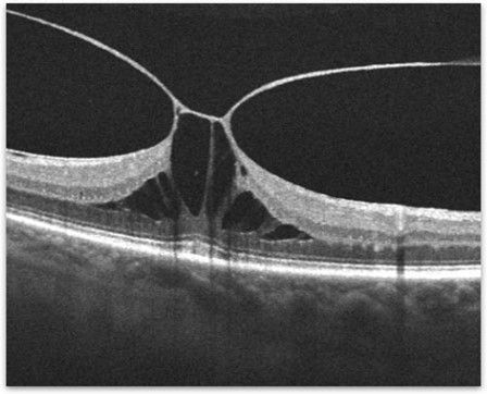
Optical coherence tomography (OCT) is the most common examination performed for the diagnosis and monitoring of patients. This is a technology that provides us with cross-sections of the retina and the vitreous surface. Thus, in great detail we can examine abnormalities on the vitreoretinal surface.

Figure 1. OCT of a patient with severe vitreous traction and cystic retinal edema.

What is the treatment for vitreous traction syndrome?

There are 3 main treatment options for vitreous traction syndrome.

  1. Careful monitoring of patients at regular intervals with OCT. This mainly concerns asymptomatic patients whose central vision has not yet been affected.
  2. For patients whose central vision has been significantly affected, the main treatment is surgery. The surgery is called vitrectomy or vitrectomy. The surgery involves the surgical removal of the vitreous through microscopic trocars (25g) in the white part of the eye (sclera). Some studies have shown that the sooner the surgery is done, the better the recovery of vision.
  3. Pneumatic vitreolysis- a small bubble of gas is injected into the eye. Then we ask the patient to look down several times an hour for 1 to 2 days. Thus, the continuous movement of the bubble within the eye releases the pull of the vitreous on the retina and macula.

Most patients with vitreous traction maintain good vision even if treatment is needed. Contact specialist ophthalmic surgeon Georgios Trichonas to learn more about your treatment options.

Intraocular lens displacement after cataract surgery and treatment

Dr.George Trichonas

Ophthalmologist Surgeon

  2111186471
 contact@retinaeyeclinic.gr

Intraocular lens displacement after cataract surgery and treatment

Often patients complain of blurred vision after cataract surgery and one of the causes is intraocular hyperdisplacement or improper positioning during surgery. Cataract surgery is a minimally invasive and immediate surgery with excellent results. In some rare cases, however, there are complications that cause vision loss and require a new corrective operation.

What happens when an intraocular lens is displaced?

Recently a patient complained to me that the lens had been dislodged from his eye. Of course, there is no such thing because the lens is immunologically neutral in the eye and is not expelled under any circumstances. Basically what is happening is that it is not resting properly inside the eye and is misaligned. A displaced intraocular lens can move into the vitreous cavity and contact the retina which is very dangerous. For this reason, the problem of displacement-movement of the intraocular lens must be treated immediately.

What could be the causes?

During cataract surgery, the clouded natural lens of the eye is removed and replaced with an artificial intraocular lens. In some cases, this extremely thin intraocular lens becomes displaced. Intraocular lens dislocation can occur days to years after surgery and may be due to trauma to the eye or diseases that affect the stability of the capsule or improper positioning during surgery.

Symptoms during intraocular displacement

Patients with intraocular lens displacement may experience a decrease or change in vision, diplopia, and sensitivity to bright light. In addition, they may report eye pain or headaches and inflammation. Some patients also report seeing the tip of the intraocular lens.

How is intraocular lens displacement treated?

There are various surgical treatments. The most appropriate with the least complications is the placement of a scleral support lens. The lens is usually sutured to the sclera (white of the eye) with a special goretex or prolene suture. Recently, a scleral support intraocular lens was released in Greece, which does not need sutures for its support. The lens is called Carlevale and with special “legs” it rests on the scleral wall of the eye, without stitches. Also, this lens presents excellent stability to avoid refractions. This lens to date has been used by many surgeons successfully and tends to be established as the lens of choice in such select cases.

With prompt and careful management, most patients with intraocular misalignment have an excellent visual outcome following the corrective procedure. Contact your specialist eye surgeon Georgios Trichona for an immediate diagnosis and treatment of the problem.

What does retinal surgeon mean?

Dr.George Trichonas

Ophthalmologist Surgeon

  2111186471
 contact@retinaeyeclinic.gr

What does retinal surgeon mean?

A retinal surgeon is a physician who has specialized in ophthalmology and then sub-specialized in diseases of the vitreous and retina. The retina surgeon is very specialized. He is a graduate of medical school, he has done a specialty in ophthalmology during which he mainly specializes in cataract surgery and then sub-specializes in retinal diseases and his surgeries. This includes:
  • Medical school – 6 years
  • Ophthalmology specialty – 4 years
  • Retina Specialization – 2 years
Most retina specialists focus their practice exclusively on the diagnosis and treatment of retinal conditions and diseases. They usually see patients who have been referred by a general ophthalmologist who has identified a potentially serious disease of the retina. Retina specialists are trained and skilled in using advanced tools and equipment to diagnose problems.

What cases does the retinal surgeon handle?

The retina surgeon can diagnose diseases of the vitreous and retina through a detailed clinical examination with special lenses and using diagnostic machines. Retina surgeons work on a very sensitive and delicate body tissue, the retina. The retina is located inside the eye and is accessible with special tools that are inserted into the eye with small trocars. Microscopes and lasers are used by the doctor very often both in the clinic and in the operating room and are vital for the treatment of retinal diseases. Specialist ophthalmologists treat a wide range of retinal diseases from age-related macular degeneration, vein occlusion and diabetic retinopathy to surgical cases such as retinal detachment, membrane and retinal hole.

Ophthalmologist Georgios Trihonas

Trichonas George, is an ophthalmologist surgeon, specialized in vitreoretinal diseases. The physician has been an assistant professor of ophthalmology at Case Western Reserve University and director of the retina department at University Hospitals, Cleveland Medical Center, USA. The doctor has also specialized in ophthalmology at the Cleveland Clinic Foundation (CCF) and is a graduate of the American Academy of Ophthalmology (American Board of Ophthalmology-ABO-Certified). Dr. Georgios Trichonas is an internationally recognized retinal surgeon. From his many years of experience abroad, he is uniquely qualified to diagnose and treat problems affecting the back of the eye, including diabetic retinopathy, macular degeneration and retinal detachment. Contact the specialized eye surgeon for a personalized diagnosis and treatment of your problem.

Retinal tear

Dr.George Trichonas

Ophthalmologist Surgeon

  2111186471
 contact@retinaeyeclinic.gr

Retinal tear 

Retinal tear is a condition that must be treated immediately by a specialist ophthalmologist. This is a tear in the retina, which is a light-sensitive tissue (like photographic film) inside the eye. This crack is very dangerous as it can cause retinal detachment, a condition that leads to blindness.

Figure 1.- OCT of retinal tear with hemorrhage.

What are the symptoms of retinal tear?

Symptoms are usually flashes and many flies as if seeing through a fog. The most dangerous symptom is when we have a deficit in our visual field like a curtain. Then usually the crack has caused a retinal detachment.

If you have any of the above symptoms, contact your specialist ophthalmologist immediately. Until you undergo a fundoscopy check it is important to stay calm, avoid heavy lifting, sudden movements and bending over.

What are the predisposing factors that cause a retinal tear?

Predisposing factors are mainly high myopia and age over 50 years. It is usually due to traction on the vitreous during vitreous detachment.

How is a retinal tear treated?

The crack must be immediately entrenched using Argon Laser photocoagulation.

In our clinic you will find Argon laser from the company Lumenis which has the upgraded Laser application with the indirect ophthalmoscope (LIO) method. This is the most painless and modern method where the patient now lying on an examination chair is subjected to treatment without the use of a lens that comes into contact with the eye. The treatment is done only with anesthetic drops and the entrenchment is done up to the extreme circumference with the help of scleral pressure when needed.

Figure 2.- OCT of the same crack 1 week after Argon Laser photocoagulation

What to expect after Argon Laser photocoagulation?

The eye may be irritated and red for a few days after the Laser. Recovery is very quick and the patient can return to work the next day with mild restrictions such as avoiding heavy lifting and vigorous activity.

The clinic of the specialized ophthalmologist surgeon Georgios Trichonas is fully equipped with diagnostics state-of-the-art machinery. It has all the necessary equipment for a complete and thorough ophthalmological examination and at the same time for the performance of all modern Lasers.

Hyalonectomy

Dr.George Trichonas

Ophthalmologist Surgeon

  2111186471
 contact@retinaeyeclinic.gr

Vitrectomy 

What is vitrectomy (vitrectomy)?

Vitrectomy or vitrectomy is a surgical procedure in which the vitreous is removed from the inside of the eye. The vitreous is often responsible for retinal diseases and its removal leads to retinal healing. The retina is a light-sensitive tissue located at the back of the eye. The vitreous is a clear jelly that fills the inside of the eye.

Why might someone need a vitrectomy?

  • Retinal detachment.
  • Severe eye injury.
  • Membrane with retinal wrinkles or otherwise epi-retinal membrane.
  • Macular hole.
  • Problems after cataract surgery.
  • Internal bleeding in the eye mainly due to diabetic retinopathy.

How is vitrectomy performed and how can it improve our vision?

According to vitrectomy special instruments of 25, 23 or 27 G are used with which special manipulations are made inside the eye. Through small trocars placed on the sclera (white part of the eye), forceps are inserted into the eye with which scar tissue or some membrane that distorts the retina can be removed. Our vision is blurred when the retina is distorted. Also a foreign body can become lodged in the eye after trauma. Most foreign bodies cause vision loss if not removed in time. Vitrectomy often improves or stabilizes our vision. During the surgery, all opacities are removed from the inside of the eye, whether from bleeding or inflammation or infection, so that light can focus on the retina. During surgery, the vitreous is replaced by a liquid that looks like salt water.

What is the vitrectomy surgery procedure?

Preoperative

Before the surgery you will need to do some general blood tests and a cardiogram. If it is a general anesthesia, a chest X-ray will also be needed. Surgery is usually performed under local anesthesia and sedation.

Interoperative
The surgery can last one or more hours depending on the condition of the eye. The surgery is performed using a microscope. The manipulations inside the eye are done using a trocar cannula that is tiny and the incisions are made in the sclera, which is the white part of the eye. During surgery your ophthalmologist may laser the retina to reduce the chances of future bleeding or to treat a retinal tear. At the end of the surgery, an air or gas bubble is often placed inside the eye to attach the retina. The bubble will be absorbed slowly. In selected cases it may place silicone oil which will need to be removed in the future.

After surgery

The vitrectomy is now in most cases done without the use of sutures, with the result that the recovery of the patients is very fast. After the surgery the eye will remain closed until the next day when you will be examined by your ophthalmologist. There may be some tenderness, foreign body sensation and mild pain in the eye. The eye will need to be covered with some gauze. Your eye doctor will prescribe some eye drops and advise you about your daily activities. If a gas bubble is placed inside the eye your ophthalmologist will tell you if a specific position of the head is needed until it is absorbed. Do not fly by plane or climb to a high altitude until the bubble is absorbed. A sudden change in altitude causes a dangerous increase in intraocular pressure with disastrous consequences.


What are the risks associated with the surgery?

  • All surgeries have some risks, but the risk is far less than the benefit that asurgery. The most common side effect of vitrectomy is a cataract, which usually occurs months after surgery.

Some of the other risks may be:

  • Retinal detachment, uncommon.
  • Reduced vision, not common.
  • High pressure in the eye, not common.
  • Infection, rare.
  • Bleeding, rarely.

How much will vision improve after vitrectomy surgery?

Your vision after surgery depends on many factors, but mainly what caused your vision to decrease before surgery. Macular lesions or macular detachment are major aggravating factors. Your eye doctor will discuss your prognosis with you before surgery.

Flies and the dangers they hide

Dr.George Trichonas

Ophthalmologist Surgeon

  2111186471
 contact@retinaeyeclinic.gr

Flies and the dangers they hide

At some point in our lives, all of us have seen flies pass before our eyes. But what do we mean by “flies”? They may look like small spots, dots, circles, semicircles, lines or spider webs, which appear in our field of vision. While they appear to be in front of us, they are actually floating inside the eye. They occur when the vitreous, a jelly-like substance that fills the eye, begins to shrink and liquefy, creating a shadow on the retina. That is, the visual impression of spots or thin lines that “float” freely within the field of vision is created for the patient.

What can they be caused by?

Flies usually appear when looking at something white or very bright. They are usually part of a normal aging process, but sometimes they can hide something more serious like a retinal tear or inflammation inside the eye.

Risk factors include:

  • Age 45 years or older
  • Refractive problems, such as mainly high myopia
  • After cataract surgery
  • After trauma
  • Inflammation in the eye

Do the flashes mean that?

The retina of the eye works like the film of a camera. That is, it receives a light stimulus and then translates it into an electrical stimulus, which is sent to the brain and creates visual perception. So when the retina has undergone some kind of tear, the vision cells receive mechanical stimulation and send an electrical stimulus to the brain that is perceived by the patient as a “flash”. Flashes and flashes may look like flashing lights or lightning in your field of vision. Flashes may occur in one or both eyes.

When should you visit the eye doctor?

When flies appear at the same time as intense flashes it can be a symptom:

In this case, the need to make an appointment with a specialized ophthalmologist becomes imperative as a fundoscopy must be performed. Underestimation of the problem and inadequate treatment can lead to vision loss. Contact the specialized ophthalmologist Georgios Trichona in vitreous-retinal diseases for a personalized diagnosis, recommending a specific treatment plan, based on the stage of the disease and your symptoms.

Ηλικιακή Εκφύλιση Ωχράς Κηλίδας και Διατροφή

Dr.George Trichonas

Ophthalmologist Surgeon

  2111186471
 contact@retinaeyeclinic.gr

Age-related Macular Degeneration and Nutrition

The macular degeneration is one of the consequences of aging. However, a healthy lifestyle can significantly reduce the risk of eye health problems. A healthy diet plays a very important role in age-related eye diseases and can reduce the risk of them by 25%.

What is age-related macular degeneration and what are the main causes of its appearance?

Age-related macular degeneration is an especially common eye condition that occurs after the age of 50. In particular, the condition is characterized by changes that occur in the macula, that is, an area at the back of the eye, in the center of the retina, that allows us to clearly distinguish the details of an image. The main causes of its appearance are age, genetic factors, smoking and diet.

Macula and nutrition: Nutrients that prevent it

Besides age, the next risk factor is poor diet. Studies conducted by the National Eye Institute have shown that there are some nutrients or a combination of them that protect against the progression of the condition.

Vitamin A

Vitamin A plays a crucial role in your vision. Vitamin A deficiency is rare in developed countries, but if present and untreated it can lead to serious health conditions. Some studies show that diets high in vitamin A are associated with a reduced risk of macular degeneration and cataracts. Foods rich in vitamin A are sweet potatoes, liver, leafy green vegetables, carrots, tomatoes, pumpkins, red peppers, etc.

Vitamin E

Many eye conditions are believed to be related to oxidative stress, which is an imbalance between antioxidants and free radicals in the body. Vitamin E is a powerful antioxidant that helps protect cells, including eye cells. Some options rich in vitamin E are nuts, seeds and cooking oils. Salmon, avocado and green leafy vegetables are also good sources.

Vitamin C

Like vitamin E, vitamin C is a powerful antioxidant that can protect your eyes from harmful free radicals. Studies have shown that when taken daily it can reduce the risk of macular degeneration by 25%. Citrus and tropical fruits, peppers, broccoli and cabbage contain particularly high amounts of vitamin C, making them excellent choices.

Lutein and Zeaxanthin

Lutein and zeaxanthin are part of the carotenoid family, a group of beneficial compounds synthesized by plants. Both of these carotenoids are believed to be able to limit oxidative damage to the retina with the help of light. A daily intake of 6 mg from a diet rich in fruits and vegetables is recommended. Green leafy vegetables such as spinach are also a good source.

Omega-3 fatty acids

Omega-3 fatty acids are a type of polyunsaturated fat and offer a neuroprotective effect on the retina, as they are found in large amounts in this tissue. In addition to helping shape your eye cells, these fats have anti-inflammatory properties that may play a role in preventing many eye conditions. To increase omega-3 fatty acids in your diet, include rich sources such as fish, flaxseed, chia seeds, soy and nuts. Omega-3s can also be found in cooking oils such as olive oil.

Research shows that certain vitamins and nutrients can help prevent or slow the progression of many different eye conditions. Supplements can be beneficial if you suspect that some of these vitamins are lacking in your diet. However, eating a balanced diet rich in fruits, vegetables, whole grains, protein and healthy fats will give you all the nutrients your eyes and the rest of your body need.

Cataract Surgery and Intraocular Lenses

Dr.George Trichonas

Ophthalmologist Surgeon

  2111186471
 contact@retinaeyeclinic.gr

Cataract Surgery and Intraocular Lenses 

waterfall is the clouding of the lens of the eye that causes a decrease in vision. It usually occurs in people over 50 years of age. During cataract surgery, the clouded natural lens of the eye is removed and replaced with an artificial intraocular lens. Improvements in technology and ever-evolving cataract removal techniques give the doctor and patient the option to treat refractive problems such as myopia, presbyopia, farsightedness and astigmatism in addition to removing the lens and treating the cataract.

Before the operation, the ophthalmologist will examine all the options and select the appropriate intraocular lenses that will be personalized to the patient’s needs.

What are the different types of intraocular lenses?

Now there is a variety of intraocular lenses that are personalized to the needs of each patient.

Monofocal Lenses

A monofocal lens is the most common type of intraocular lens used in cataract surgery. This type of lens is designed to give you sharp and clear vision at a certain distance. Some patients choose a monofocal lens that focuses well on objects 45 to 90 centimeters away from the face. This is the distance at which most computers are placed, and this lens choice allows them to do their work every day without glasses. Others who enjoy reading choose a monofocal lens that offers near vision.

Multifocal Intraoculars

The multifocal lens is a multifocal intraocular lens designed to correct presbyopia and provide a full range of near, far, and intermediate vision along with improved image quality. After a study it was found that people with multifocal lenses had more sensitivity to light, which makes it difficult to drive at night as the lights from running cars can cause discomfort to the patient.

Toric Intraocular Lenses

Toric intraocular lenses are aimed at patients with astigmatism, thus freeing them from wearing glasses. In a published article, they interviewed eye doctors and asked them what kind of intraocular lenses they would recommend for patients with astigmatism. Among respondents, 48% chose a toric lens, making this the most popular type of lens for those with astigmatism.

The Monovision Method

In the monovision method, each eye is adjusted so that it can focus at a different distance. That is, it is the technique in which one eye is corrected for distant vision and the other for near vision. It is indicated for patients who cannot be implanted with multifocal intraocular lenses. 80% of people in the general population who suffer from cataracts can be treated with the monovision method.

Which type of lenses is right for you?

Every patient’s daily life and needs are different. Some factors that will help you choose the right intraocular lenses, always with the cooperation of the ophthalmologist, are:

  • Daily life and lifestyle
  • Night driving
  • Degree of astigmatism
  • Other eye diseases
  • Cost of intraocular lenses

Your doctor can assess how healthy your eyes are and can help you understand how each lens can work to help you achieve your goals.

ΚΑΛΕΣΤΕ ΜΑΣ